Congenital Hip Dislocation: Timely Identification of the Problem
Содержимое
Early detection and diagnosis of congenital hip dislocation is crucial for effective treatment and prevention of long-term complications. This article provides information on the importance of timely identification and intervention for this common childhood condition.
Congenital hip dislocation is a condition that affects infants and can lead to long-term mobility issues if not detected and treated early. The hip joint is a crucial part of the body’s skeletal structure, allowing for smooth movement and stability. When the hip joint is not properly formed or positioned at birth, it can result in hip dislocation.
Early detection of congenital hip dislocation is essential for effective treatment and prevention of complications. Newborns are routinely screened for this condition through physical examination techniques, such as the Ortolani test or Barlow maneuver. These tests involve gently manipulating the baby’s legs to check for any signs of instability or dislocation in the hip joints.
It is important for parents and caregivers to be aware of the risk factors associated with congenital hip dislocation. These include a family history of the condition, breech presentation during pregnancy, and certain fetal positioning during development. By being aware of these risk factors, parents can work closely with healthcare professionals to ensure early detection and intervention if necessary.
Early detection and prompt treatment of congenital hip dislocation can greatly improve outcomes for children affected by this condition. Treatment options may include the use of a Pavlik harness, which helps to position the hip joint correctly and promote proper development. In some cases, surgical intervention may be required to correct the hip joint’s alignment.
Overall, early detection of congenital hip dislocation is crucial for the long-term health and mobility of infants. By being aware of the risk factors and actively participating in screening processes, parents can ensure that their child receives the necessary care and intervention to optimize their hip joint function.
Congenital Hip Dislocation

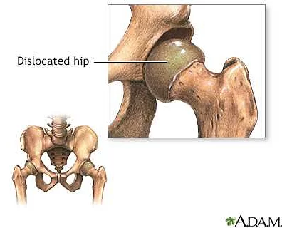
Congenital hip dislocation, also known as developmental dysplasia of the hip (DDH), is a condition in which the hip joint is not properly formed in a newborn baby. This results in the ball of the thighbone (femur) not fitting correctly into the socket of the hipbone (acetabulum).
DDH can range from mild to severe, with some cases resolving on their own while others requiring treatment. If left untreated, it can lead to long-term problems such as pain, difficulty walking, and hip joint instability.
Early detection is crucial in managing congenital hip dislocation. The condition can often be diagnosed during a routine physical examination in newborns. Doctors will look for signs such as an uneven leg length, limited hip movement, and a clicking sound in the hip joint.
It is recommended that all newborns undergo a hip examination within the first few days of life. This can be done by a healthcare provider who is trained in detecting developmental dysplasia of the hip. If any signs or risk factors are present, further diagnostic tests such as ultrasound or X-rays may be recommended.
Treatment for congenital hip dislocation depends on the severity of the condition. In mild cases, the hip may improve on its own with simple measures such as using a special harness or brace to keep the hip in the correct position. In more severe cases, surgery may be required to reposition the hip joint and ensure proper development.
Regular follow-up visits with a healthcare provider are important to monitor the progress of treatment and ensure that the hip is developing normally. With early detection and appropriate treatment, most cases of congenital hip dislocation can be successfully managed, allowing for normal hip development and function.
Understanding the Condition

Congenital hip dislocation is a condition that affects infants. It occurs when the hip joint is not properly formed, causing the head of the femur to be partially or completely dislocated from the hip socket. This condition can range from mild to severe, and if left untreated, it can lead to long-term complications and affect the child’s ability to walk.
There are several factors that can contribute to the development of congenital hip dislocation. These include a family history of the condition, breech position during pregnancy, first-born child, and female gender. It is important for parents and healthcare providers to be aware of these risk factors and monitor infants for any signs or symptoms of the condition.
Early detection of congenital hip dislocation is crucial for successful treatment. Regular physical examinations and screenings are recommended, especially during the first few weeks and months of a baby’s life. During these exams, healthcare providers will look for signs such as limited hip movement, uneven leg lengths, or a clicking sound when moving the hip joint.
If congenital hip dislocation is suspected, further diagnostic tests may be done, such as an ultrasound or an X-ray. These tests can help determine the severity of the condition and guide treatment options. Treatment for congenital hip dislocation may involve the use of a device called a Pavlik harness, which helps to properly position the hip joint and encourage normal development. In more severe cases, surgery may be required to correct the dislocation.
Early detection and treatment of congenital hip dislocation is essential to ensure the best possible outcomes for affected infants. With appropriate care and intervention, most children with this condition are able to lead normal, active lives without long-term complications.
| – Limited hip movement – Uneven leg lengths – Clicking sound when moving the hip joint | – Family history of the condition – Breech position during pregnancy – First-born child – Female gender |
Signs and Symptoms

Congenital hip dislocation is a condition that affects the development of the hip joint in newborns. It can cause pain and discomfort and may lead to difficulty with walking later in life if left untreated. There are several signs and symptoms that may indicate the presence of congenital hip dislocation:
1. Limited range of motion: One of the first signs of congenital hip dislocation is limited movement in the affected hip joint. The infant may have difficulty fully extending or flexing the hip, and the range of motion may be noticeably less than in the unaffected hip.
2. Abnormal hip position: Another common sign is an abnormal positioning of the hip joint. The affected hip may be dislocated out of its socket, resulting in a visible asymmetry or unevenness in the hips. The affected leg may appear shorter or rotated compared to the unaffected leg.
3. Clicking sound: A clicking sound may be heard when the hip joint is moved. This sound is caused by the abnormal movement of the femoral head in the hip socket. It is important to note that not all infants with hip dysplasia will experience this clicking sound.
4. Uneven skin folds: Another potential sign is uneven skin folds on the buttocks or thighs. In some cases, the affected hip may cause the skin to fold or crease differently compared to the unaffected side.
5. Delayed walking: In more severe cases, children with congenital hip dislocation may experience a delay in walking. This is because the abnormal hip joint can affect the child’s ability to bear weight and balance properly. Parents should consult with a healthcare professional if their child is not walking by the expected age.
If any of these signs or symptoms are present, it is important to seek medical attention for further evaluation and diagnosis. Early detection and intervention are crucial for a successful treatment outcome.
Note: It is essential to consult with a healthcare professional for a proper diagnosis and treatment plan. This article is for informational purposes only and should not be used as a substitute for professional medical advice.
Diagnosis and Screening Methods
Diagnosing congenital hip dislocation requires a combination of physical examination and imaging tests. Early detection is crucial in order to prevent long-term complications and to ensure timely treatment.
During the physical examination, the healthcare provider will assess the range of motion in the baby’s hips and perform a series of maneuvers to check for instability. These maneuvers may include the Barlow test, in which the provider gently pushes the baby’s hip out of its socket, and the Ortolani test, in which the provider gently moves the hip back into its socket.
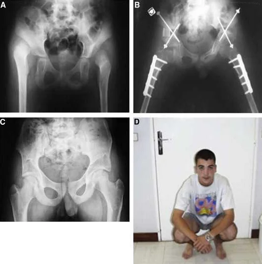
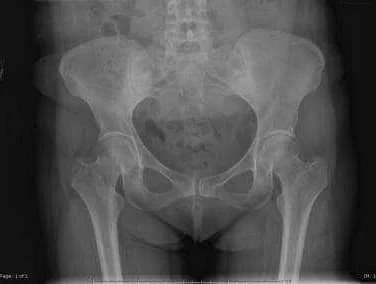
In addition to the physical examination, imaging tests such as ultrasound and X-rays are commonly used to confirm the diagnosis and determine the severity of the condition. Ultrasound is a safe and non-invasive imaging tool that can visualize the hip joint and identify any abnormal positioning or displacement of the hip. X-rays can provide more detailed information about the hip joint and are useful in assessing the development of the bones.
Screening methods for congenital hip dislocation are crucial in identifying the condition early, even before symptoms become apparent. Many countries have implemented universal newborn screening programs, in which all newborns undergo a physical examination and imaging tests within the first few weeks of life. This helps to identify infants with hip dysplasia or dislocation and allows for early intervention and treatment.
In conclusion, the diagnosis of congenital hip dislocation involves a combination of physical examination and imaging tests. Early screening is essential in order to detect the condition before symptoms develop, allowing for timely intervention and treatment.